Spectral Karyotyping: Advanced Chromosome Analysis in Modern Genetic Research

Genetic research and molecular diagnostics have transformed modern healthcare by improving the understanding of chromosomal abnormalities and genetic disorders. One of the most advanced techniques used in cytogenetics today is spectral karyotyping.
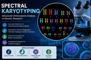
Spectral karyotyping is a powerful imaging technology that allows scientists and healthcare professionals to visualize chromosomes in multiple colors for accurate genetic analysis. This advanced method has become highly valuable in cancer research, prenatal diagnostics, genetic disorder analysis, and biomedical research.
With the integration of advanced imaging systems, cytogenetics software, and biotechnology tools, spectral karyotyping is helping researchers identify complex chromosomal changes with greater precision and efficiency.
In this article, we will explore what spectral karyotyping is, how it works, its applications, advantages, and its growing importance in modern genetic diagnostics.
What Is Spectral Karyotyping?
Spectral karyotyping is an advanced cytogenetic imaging technique used to analyze chromosomes by labeling each chromosome pair with different fluorescent colors.
This technique helps scientists identify:
- Chromosomal abnormalities
- Genetic rearrangements
- Translocations
- Duplications
- Deletions
- Complex chromosome structures
Unlike traditional chromosome analysis methods, spectral karyotyping provides highly detailed visualization, making it easier to detect genetic abnormalities accurately.
What Is Karyotyping?
To understand spectral karyotyping, it is important to know the basics of karyotyping.
Karyotyping is a laboratory technique used to examine the number, size, and structure of chromosomes in a cell.
Standard karyotyping helps diagnose:
- Genetic disorders
- Birth defects
- Infertility issues
- Certain cancers
- Chromosomal abnormalities
However, traditional karyotyping may have limitations in identifying complex chromosomal changes. Spectral karyotyping overcomes these limitations through advanced fluorescent imaging technologies.
How Spectral Karyotyping Works
Spectral karyotyping uses specialized fluorescent probes that bind to chromosomes. Each chromosome pair is labeled with a unique combination of fluorescent dyes.
The process typically includes:
- Cell sample preparation
- Chromosome isolation
- Fluorescent probe application
- Imaging using advanced microscopy systems
- Chromosome analysis through cytogenetics software
The resulting image displays chromosomes in multiple colors, allowing researchers to identify abnormalities more efficiently.
Importance of Spectral Karyotyping in Genetic Research
Spectral karyotyping has become highly important in modern biomedical and genetic research.
Key Benefits of Spectral Karyotyping
- Improved chromosome visualization
- Accurate identification of genetic abnormalities
- Detection of complex chromosomal rearrangements
- Enhanced cancer research capabilities
- Faster and more precise analysis
Advanced imaging systems combined with karyotyping reagents improve laboratory efficiency and diagnostic accuracy.
Applications of Spectral Karyotyping
Spectral karyotyping is widely used across several healthcare and research applications.
Cancer Diagnostics and Research
Cancer Diagnostics cells often contain abnormal chromosome structures and genetic rearrangements.
Spectral karyotyping helps researchers identify:
- Leukemia-associated abnormalities
- Lymphoma chromosomal changes
- Tumor genetics
- Cancer progression markers
Modern cancer diagnostics increasingly rely on advanced cytogenetics technologies for accurate genetic analysis.
Prenatal Genetic Testing
Spectral karyotyping supports prenatal screening for chromosomal abnormalities that may affect fetal development.
It helps detect conditions such as:
- Down syndrome
- Turner syndrome
- Chromosomal duplications
- Structural rearrangements
Genetic Disorder Analysis
Researchers use spectral karyotyping to study inherited genetic disorders and chromosomal diseases.
This technique provides valuable insights into rare genetic abnormalities and developmental conditions.
Biomedical Research
Biotechnology companies and research laboratories use spectral karyotyping for:
- Cell biology research
- Gene mapping
- Chromosome studies
- Molecular diagnostics
Role of Karyotyping Reagents in Chromosome Analysis
Karyotyping reagents are essential for preparing chromosome samples and enabling fluorescent labeling.
These reagents help improve:
- Chromosome staining quality
- Fluorescent signal accuracy
- Imaging clarity
- Diagnostic consistency
High-quality karyotyping reagents are critical for obtaining reliable chromosome analysis results.
Cytogenetics Software and Imaging Systems
Modern spectral karyotyping systems integrate advanced imaging technologies with specialized cytogenetics software.
These systems support:
- Automated chromosome analysis
- Image enhancement
- Data interpretation
- Digital reporting
- Real-time visualization
Digital cytogenetics platforms significantly improve laboratory workflow and research productivity.
Importance of Spectral Karyotyping in Oncology
Oncology research heavily depends on chromosome analysis because many cancers are linked to genetic mutations and chromosomal abnormalities.
Spectral karyotyping helps oncologists and researchers:
- Understand tumor genetics
- Identify treatment targets
- Monitor cancer progression
- Improve personalized medicine strategies
Advanced chromosome imaging contributes to more accurate cancer diagnosis and treatment planning.
Advantages of Spectral Karyotyping Over Traditional Methods
Spectral karyotyping offers several advantages compared to conventional chromosome analysis techniques.
Higher Accuracy
Multiple fluorescent colors improve chromosome differentiation and abnormality detection.
Better Visualization
Complex chromosome rearrangements become easier to identify.
Faster Analysis
Automated imaging and software systems improve laboratory efficiency.
Enhanced Research Capabilities
Researchers can study complex genetic interactions more effectively.
Challenges in Spectral Karyotyping
Despite its advantages, spectral karyotyping also faces certain challenges.
High Equipment Costs
Advanced imaging systems and fluorescence microscopes require significant investment.
Technical Expertise Requirements
Proper chromosome analysis requires trained cytogenetic specialists.
Complex Data Interpretation
Chromosomal analysis may involve complicated genetic patterns requiring advanced software and expertise.
Sample Preparation Sensitivity
Accurate results depend heavily on proper laboratory procedures and sample quality.
Future of Spectral Karyotyping
The future of spectral karyotyping is closely connected with advancements in biotechnology, artificial intelligence, and digital imaging systems.
Emerging developments include:
- AI-powered chromosome analysis
- Automated cytogenetics platforms
- High-resolution imaging systems
- Integrated genomic analysis
- Cloud-based laboratory data management
These innovations will continue improving diagnostic precision and genetic research efficiency.
Spectral Karyotyping and Precision Medicine
Precision medicine focuses on personalized healthcare based on individual genetic profiles.
Spectral karyotyping contributes to precision medicine by helping doctors:
- Identify genetic mutations
- Understand disease mechanisms
- Select targeted therapies
- Monitor treatment responses
Advanced chromosome analysis technologies are becoming increasingly important in personalized healthcare and cancer treatment.
Conclusion
Spectral karyotyping is one of the most advanced chromosome analysis techniques used in modern cytogenetics and genetic research.
By combining fluorescent imaging, advanced microscopy, and cytogenetics software, spectral karyotyping enables accurate detection of chromosomal abnormalities and genetic rearrangements.
From cancer diagnostics and prenatal screening to biomedical research and precision medicine, spectral karyotyping continues to improve scientific understanding and healthcare outcomes.
As biotechnology and digital imaging technologies evolve, spectral karyotyping will remain a critical tool in genetic diagnostics and advanced medical research.
FAQs
What is spectral karyotyping?
Spectral karyotyping is an advanced chromosome imaging technique that uses fluorescent colors to identify chromosomal abnormalities and genetic rearrangements.
What is the purpose of karyotyping?
Karyotyping helps analyze chromosome structure, number, and abnormalities for diagnosing genetic disorders and cancers.
How is spectral karyotyping different from traditional karyotyping?
Spectral karyotyping uses multiple fluorescent dyes to improve chromosome visualization and detect complex chromosomal changes more accurately.
What are karyotyping reagents?
Karyotyping reagents are laboratory chemicals and fluorescent probes used to prepare and stain chromosomes for analysis.
Where is spectral karyotyping used?
It is widely used in cancer diagnostics, prenatal testing, genetic disorder analysis, biomedical research, and cytogenetics laboratories.
Why is spectral karyotyping important in cancer research?
Many cancers involve chromosomal abnormalities. Spectral karyotyping helps researchers identify genetic changes linked to tumor development and progression.
What role does cytogenetics software play in spectral karyotyping?
Cytogenetics software supports chromosome imaging, analysis, data interpretation, and digital reporting.
What is the future of spectral karyotyping?
The future includes AI-powered chromosome analysis, automated imaging systems, and integrated genomic diagnostics for precision medicine.